Biology
ANIMAL TISSUES
TISSUES:- Group of cells that work together to perform a particular function
ANIMAL TISSUES
Animal Tissues are more complex rather than that of plants. Its reason goes to this view that the activities of animals are much advanced in comparison to plants. For example, there is no respiration, no digestion, no movement in plants. but these are carried out by animals. Thus they need some special systems in their body to perform these functions. These systems are produced by the interaction of certain specific groups of cells, these groups are known as Animal Tissues.
Animal Tissues are classified as follows:-
Covering Epithelia:- The tissues that form the covering of outer and inner surfaces of various organs of the body is called covering epithelia. Thus tissues generally serve as a protective covering both internally and externally. Since they cover the surface of the body or line the internal organs they are also called ‘boundary tissues’.
The cells of this tissue are highly packed and form a continuous sheet. Inter-cellular matrix (Interstitial fluid) very little or absent. Epithelial cells lie on a delicate non-cellular basement membrane. The basement membrane is produced due to secretion of protein by epithelial cells and association of it with mucopolysaccharides (Hyaluronic acid and glycoproteins). The reticular fibers also take part in its synthesis. The basement membrane is permeable in nature due to which the chemical exchange between epithelial cells and the tissue fluid of connective tissue takes place through it.
Functions of Epithelia
- These form the outer layer of skin. These protect the underlying cells of from drying, injury and chemical effects. They also protect the body from viral or bacterial infection.
- They help in chemical and gas exchange between internal organs along with body and outer environment.
- They help with sensory reception.
- The living of mouth and alimentary canal and protection of organs.
- Help in the elimination of waste products.
- Absorption of water and nutrients.
- They perform secretion, they secrete, sweat, saliva, enzymes etc.
- They perform regeneration.
TYPES OF EPITHELIA
(A) SQUAMOUS EPITHELIA (Squama = Scale)
Structure:- It is made up of thin, plat like (flat) polygons which fit together like mosaic files. Thus also called Pavement Epithelia.
Occurrence:- Present on the lining of body and internal organs, it forms the coelomic epithelium or peritoneum which is called mesothelium. The covering of heart, blood and lymph capillaries is also squamous epithelia. It is called Endothelium.
Except above squamous epithelia is also present in terminal bronchioles, Air chambers. Bowman’s
Capsules of kidneys, Henle’s loop canal and in Testies (forming rete testis).
Note:- Cells of Mesothelia and Endothelia are interconnected with each other with the help of finger-like interdigitations. So these epithelia are called tessellated.
Functions:-
- Protection of underlying parts of the body from mechanical injury, entry of germs, chemicals, and drying.
- It makes the surface selectively permeable due to which chemical exchange takes place.
- Conduction of fluid materials in capillaries, absorption excretion and as sensory.
(b) Cuboidal epithelium
Structure:- It consists of cube-like cells which appears square in section nucleus is very distinct and spherical, thyroid vesicles, ciliary body and choroid of eyes and in their respiratory ducts. Also present in some glands (thyroid, sweat glands, pancreas etc) and in germinal epithelium of gonads (Testies and ovaries).
Function:- Absorption, Excretion and Secretion and also provides mechanical support.
(c) Columnar Epithelia
Structure:- These tissues have elongated and prismatic (rectangular) cells. The nucleus is also elongated and situated at the base. In between the epithelial cells, the goblet cells are present which secrete Mucous. Sometimes free ends epithelia contain microvilli.
Occurrence:- Present in the lining of the stomach, small intestine, and colon where it forms the mucous membrane, also form the lining of gallbladder and oviducts.
Functions:- Main function is absorption (stomach, intestine) and secretion (mucous by goblet cells).
Modified Epithelia
(a) Glandular Epithelia:- Although all living cells perform secretion, the cells which are specialized to perform this function are called secretory or glandular cells. This tissue is known as glandular tissue. These cells are generally cuboidal or global cells. This tissue is also known as a gland.
(b) Ciliated Epithelia:- However some cuboidal or columnar cells have cilia on its surfaces. The tissues are also known as Ciliated Epithelia.
This tissue is found in sperm ducts. It lines the trachea, bronchi, kidney tubules and oviducts. The Cillia present on this tissue help in moving solid particles through ducts.
- Muscular Tissue
This tissue helps in the movement of body organs (internal and external). The cells of this tissue are elongated large sized so-called muscle fibers. The muscle fibers are arranged parallelly to each other so that they can work together much effectively.
This tissue is of following three types.
- Striated muscles
- Smooth muscles
- Cardiac muscles
Striated muscles
Structure:- These muscles are variably named according to their occurrence and function. As these muscle fibers have striped on them so-called striped or striated muscle fibers. As they work according to our will, these called Voluntary muscles. Due to attaching with bones, these muscles are known as skeletal muscles. These muscles help in movement of body organs (limps) and in locomotion. Thus they called somatic muscles. Also known as the phasic type of muscles as they organize rapid, but brief (short period) movements in the organs. Striated muscle fibers are 1 mm to 30 cm long, cylindrical, thread-like, unbranched and complex in structure. Around the muscles fibers, there is a three-layered covering known as sarcolemma which is very excitable due to electrically charged. The cytoplasm of muscle fibers is called sarcoplasm. It contains numerous oval-shaped nuclei. Thus cell is multinucleate. Sarcoplasm also contains hundreds of millions of elongated thread-like filaments which are arranged parallelly. These are known as myofibrils or sarcostyles. The striated nature of these muscle fibers is due to these sarcostyles.
Occurrence:- Striated muscles occur in muscles of limbs (e.g. biceps and triceps of arms), body wall, face, neck etc. Striated muscles present in tongue, pharynx, diaphragm and upper part of the esophagus are called Visceral Striated muscles.
Functions:-
- Striated muscles are powerful and undergo rapid contraction. These muscles can be tired and need rest.
- Striated muscles provide the force for locomotion and all other voluntary movements of the body.
Note:- Try to know about:-
- Muscle twitch
- Fatigue
- Paralysis
- Shivering
2. Smooth Muscles:-
These are known as unstriated, visceral or involuntary muscles. Its muscle fibers are very short (0.02 – 0.2mm length), thin and spindle-shaped and very simple in structure. Plasma membrane present on fibers but not a complete sarcolemma. Nucleus single (uninucleate), elongated and situated centrally.
Occurrence:- These muscles are found in walls of coelomic visceral organs (alimentary) canal, ducts, uterus, vagina, and blood capillaries etc) penis, eyes, dermis of skin etc. The muscle of dermis of skin are related to roots of hairs and called Arrector Pilli. Muscles of penis make reticuluous structure.
Functions:
- These muscles do not work according to our will, so they are also called involuntary muscles.
- Movement of food in the alimentary canal, opening and closing of tubes are involuntary movements.
- Due to slow contraction, these muscles show a specific characteristic movement in the alimentary canal, known as peristalsis. It includes the rhythmic progressive wave of muscular contraction and relaxation in the alimentary canal and male genital tract.
- These muscles produce extrusive movement in the urinary bladder, the gallbladder, and the uterus.
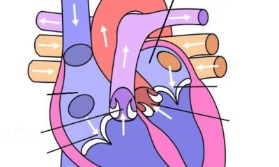
3. Cardiac Muscles:- These muscles show characteristics of both smooth and striated muscles. These composed of branched fibers, the branches form the network. The cell membrane is Sarcolemma, Cytoplasm in Sarcoplasm. It contains longitudinal myofibrils and a centrally located nucleolus (uninucleate cell). Intercellular spaces are filled with loose connective tissue supplied with blood capillaries. Cardiac muscles have stripes and also have densely stained cross-bands which are called Intercalated discs.
Occurrence:- The cardiac muscles occur in the walls of the heart.
Functions:-
- Cardiac muscles contract and relax rapidly, rhythmically and tirelessly throughout life.
- The contraction and relaxation of the heart muscles help to pump and distribute blood to various parts of the body.
Comparison of different muscles
Smooth Muscle Skeletal muscle Cardiac muscle
- Not striated Striated Striated
- Spindle – Shaped Cylindrical Cylindrical
- Not branched Not branched Branched
- Nucleus Central Nuclei –Peripheral Nuclei-Central
- No discs No discs Intercalated discs
- Involuntary Voluntary Involuntary
- Slow Fast Fast
- Contraction not inherent Contraction not inherent Contraction Inherent
CONNECTIVE TISSUE.
This is most widely distributed tissues in the body. It makes 30% part of the body. This tissue connects and anchors various body organs. The main function of this tissue is Binding, Supporting and packing together different organs of the body.
Connective tissue contains the following type of cells:-
1) Fibroblasts:– These form ground substance and fibers (e.g. Collagen, Elastin)
2) Adipose Cells:- They store fats (lipids) in their vacuoles
3) Macrophages:– These are free moving or fixed phagocytes [Leucocytes WBCS] which involve in the destruction and removal of invading bacteria, foreign bodies and damaged cells from tissues.
4) Mast Cells:- They secrete substances such as heparin (anticoagulant), Histamine (vasodilator and serotonin vaso-constructor. The promote inflammation of the infected area.
5) Immunocytes:- These include cells such as lymphocytes and plasma cells both producing antibodies for the Immune response.
The matrix of connective Tissue contains following fibers:-
1) White Fibres (made up of collagen Protein)
2) Yellow Fibres (made up of Elastic Protein)
3) Reticular Fibres (made up of Reticulin Protein)
(Reticular Fibres found mainly in growing stage)
Connective Tissue can be classified into following three categories:-
(a) Connective Tissue Proper
(b) Skeletal Tissue
(c) Vascular Tissue
(A) Connective Tissue Proper: This tissue contains delicate and jelly-like ground substance. It contains three categories:-
(1) Simple, loose connective tissues or areolar connective tissue.
(2) Dense, fibrous connective tissues
(3) Specialized connective tissues.
(1) Simple loose connective tissue structure:- It is loose and cellular connective tissues. Its matrix contains two types of fibers – White and Yellow (Collagen and Elastic), fibroblasts, plasma cells, fat cells and mast cells mainly also contain blood and lymph capillaries.
Occurrence:- It is simplest and most widely distributed connective tissues. It joins, skin to muscles, fills spaces inside organs, found around muscles, blood vessels and nerves in mesenteries and stroma.
Functions:-
- It acts as a supporting and packing tissue between organs lying in the body cavity. The matrix of this tissue is important in the diffusion of oxygen and nutrients from small blood vessels.
- It helps in repair of tissues after an injury.
- It also helps in combating foreign toxins.
- Its fixes skin to underlying muscles.
(2) Dense fibrous connective Tissue
This tissue contains mainly fibrocytes. These cells make too many fibers that all tissue become fibrous. Due to the density at fibers, this tissue is tensile. It can be categorized into two groups:-
(a) Tendons:- These contain mainly white fibers i.e. collagen. These are cord-like, collagen fibres are parallelly arranged in bundles. Also, contain fibroblasts in between bundles of fibers. These tissues are bounded by Areolar tissue.
Function:- It joins skeletal muscles to bones.
(b) Ligaments:-This tissue contains elastic fibres. It has great strength and elasticity due to elastic fibers. These are also bounded by areolar connective tissue.
Function:- It joins bones to bones. Due to strength the joints the normal movement takes place as the ligaments stretch, but sometimes elastic fibers are damaged due to overextension (overstretching). This is called strain.
(3) Specialized Connective Tissue:- This tissue is further classified to two:-
(a) Pigmented Connective Tissue:- In the dermis, choroid and Iris of eyes these are branched cells known as Chromatophores. These contain numerous black, brown, golden, white or blue pigment granules. These granules are known as Melanin. The color of our body is due to these granules. Elastic and leapt cells also present in it.
(b) Adipose or fatty connective Tissue:-
Although this tissue can form in any tissue due to modification or areolar tissue, it is specifically found the following places
(1) Below the skin of human – panniculus adiposus
(2) Blubber of whale
(3) Around the yellow bone marrow and blood vessels.
(4) Close to testes in a frog.
Note: The food reservoir of camel and thick tale of Marino sheep are due to fat storage.
Structure: Each Adipocyte is rounded or oval contains large droplets of fat that almost fills it. The fat cells are arranged into lobules separated by partitions of collagen and elastin fibers. These partitions carry blood vessels of lobules.
Functions:
1) It serves as a fat reservoir.
2) It provides shape to the limbs and the body.
3) It keeps visceral organs in position. It forms shock-absorbing cushions around kidneys and eyeballs.
4) It acts as an insulator. Being a poor conductor of heat, it reduces heat loss from the body, means it regulates body temperature.
B. Skeletal Tissue: The endoskeleton of vertebrates is made up of a specific highly dense connective tissue. It is known as skeletal tissue. It also contains a rich Inter-cellular substance. It is of two types:-
(1) Cartilage:- It is compact and less vascular. Intercellular substance i.e matrix is known as Chondrin. It contains proteins and calcium salts. Condrin is solid, cheese like and firm but also somewhat elastic. Due to which it is flexible, cells of cartilage are known as Chondrocytes which are present in fluid-filled spaces known as lacunae. Blood vessels are absent in matrix
Matrix also contains collagen fibers.
Occurrence:- Generally the endoskeleton of kids mostly contains cartilage.
Also present in ear pinna, nose-tip, epiglottis, intervertebral discs, end of long bones, lower ends of ribs and rings of the trachea.
Function:- Provides support and flexibility to the body parts. It smoothens surface at joints.
(2) Bones: Bone is very strong and non-flexible tissue. It is porous, highly vascular, mineraled, hard and rigid. Carbonates of calcium and magnesium make this tissue hard. The matrix is in the form of concentric rings called lamellae. Bone cells are called osteoblasts or osteocytes and present in fluid filled spaces lacunae which are in between lamellae. All lacunae form canaliculi. Each canaliculus is filled delicate cytoplasmic process of the bone cell. It helps in receiving food and oxygen and eliminate waste by bone.
Functions:- It serves the following functions.
1) It provides shape to the body.
2) Provides skeletal support to the body.
3) Protects organs such as brain, lungs, heart etc.
4) Serves or storage site of calcium and phosphate.
5) It anchors the muscles.
Vascular Tissue:- This tissue is also known as fluid concentric tissue. This connective tissue links the different parts of the body and maintains a continuity in the body. It includes blood and lymph.
A Blood:- The matrix of this tissue is called plasma. It contains three type of cells in it.
- Red Blood Cells (RBCs) or Erythrocytes.
- White Blood Cells, (WBCs) or Leucocytes.
- Platelets
- Red Blood Cells:- These cells have iron-containing red respiratory pigment the hemoglobin. Mostly Erythrocytes are oval or disc-shaped, nucleated and biconvex in vertebrates, but in a mammal, it is biconcave and anucleated. It is the advanced character of mammals as the cells provide more space to carry more oxygen as they transport it.
- WBcs or Leucocytes:- These cells are of two main kinds:-
1) Phagocytes
2) Immunocytes
Phagocytes are capable of phagocytosis and carry out the function of body defense by engulfing bacteria and other foreign substances.
Phagocytes are of two types:-
1) Granulocytes
2) Agranulocytes
Granulocytes have irregular- shaped nuclei and cytoplasmic granules and they are further of three types:-
1) Basophils
2) Neutrophils
3) Eosinophils
Agranulocytes have no cytoplasmic granules and include monocytes having a large nucleus and a large amount of cytoplasm. They ultimately migrate to body tissues and transform into macrophages and histiocytes.
Immunocytes produce antibodies and involved in immune response. They include lymphocytes which later on transform into plasma cells.
Blood platelets are minute anucleated, fragile fragments of giant bone marrow cells called megakaryocytes.
Occurrence. Blood occurs in blood vessels called arteries, veins, and capillaries which are connected together to form the circulatory system. The highly branching network of vessels enables blood to reach every part of the body.
Functions.
- Blood transports nutrients, hormones, and vitamins to the tissues and transports excretory products from the tissue to the liver and kidney.
- The red blood corpuscles (RBCs) carry oxygen to the tissues for the oxidation of foodstuff.
- The white blood cells (WBCs) fight disease either by engulfing and destroying foreign bodies or by producing antitoxins and antibodies that neutralize the harmful effects of germs.
- Blood platelets disintegrate at the site of injury and help in the clotting of blood.
(B) Lymph
Nature:- Lymph is a colorless fluid that has filtered out of the blood capillaries. Since it is part of blood, its composition is similar to that of blood except that red blood corpuscles and some proteins are absent in it. In the lymph, white blood cells are found in abundance.
Functions:-
- Lymph transports the nutrients (oxygen, glucose) that may have filtered out of the blood capillaries back into the heart to be recirculated in the body.
- It brings CO2 and nitrogenous wastes from tissue fluid to blood.
- Being loaded with WBCs such as lymphocytes, the lymph protects the body against infection. It forms the defense or immune system of the body.
Nervous Tissue
(which controls all the activities of the body) is composed of specialized cells, called “nerve cells” (or neurons). Each neuron is divided into two parts namely, cyton and axon.
- Cyton (also called cell body) is irregular in shape and the large nucleus is present in its cytoplasm. Each cyton has some short and branched projections. They are called the dendrons. Each Dendron divides into numerous fibers, the dendrites.
(b) Axon is a single very long, cylindrical process of uniform diameter, it arises from corner projection (axon hillock) of the cyton. The dendrites receive impulses; while the axon takes these impulses away from the cell body. In this way, neuron controls all the activities of the body.
Axon versus dendrites
| Axon |
Dendrites |
|
|